Choosing the perfect grid for your cryo-EM experiments
Imagine finding yourself, the cryogenic electron microscopy (cryo-EM) specialist, in the heart of a bustling lab, facing the critical task of preparing your sample. Your goal: unlocking the intricate details of biomolecules with near-atomic precision. The key to success lies in many factors, e.g. the purity and concentration of your sample, buffer type, and vitrification. Also choosing the perfect grid for your specific sample can be critical. This is why we will guide you through choosing the most optimal grid for your sample preparation process, shedding light on the grid selection process and help you overcome some of the challenges and optimizing your single particle research.
Selecting the right grid: A cryo-EM specialist's dilemma
As a cryo-EM specialist, you know that your grid choice can make a big impact on the experimental outcome. It's not just about picking a piece of material; it's about making an informed decision that suits the needs and goals. Which type of materials or foil pattern would you choose for example? Let's dive into the key considerations to help you choose wisely.
Grid material: Material type and mesh size
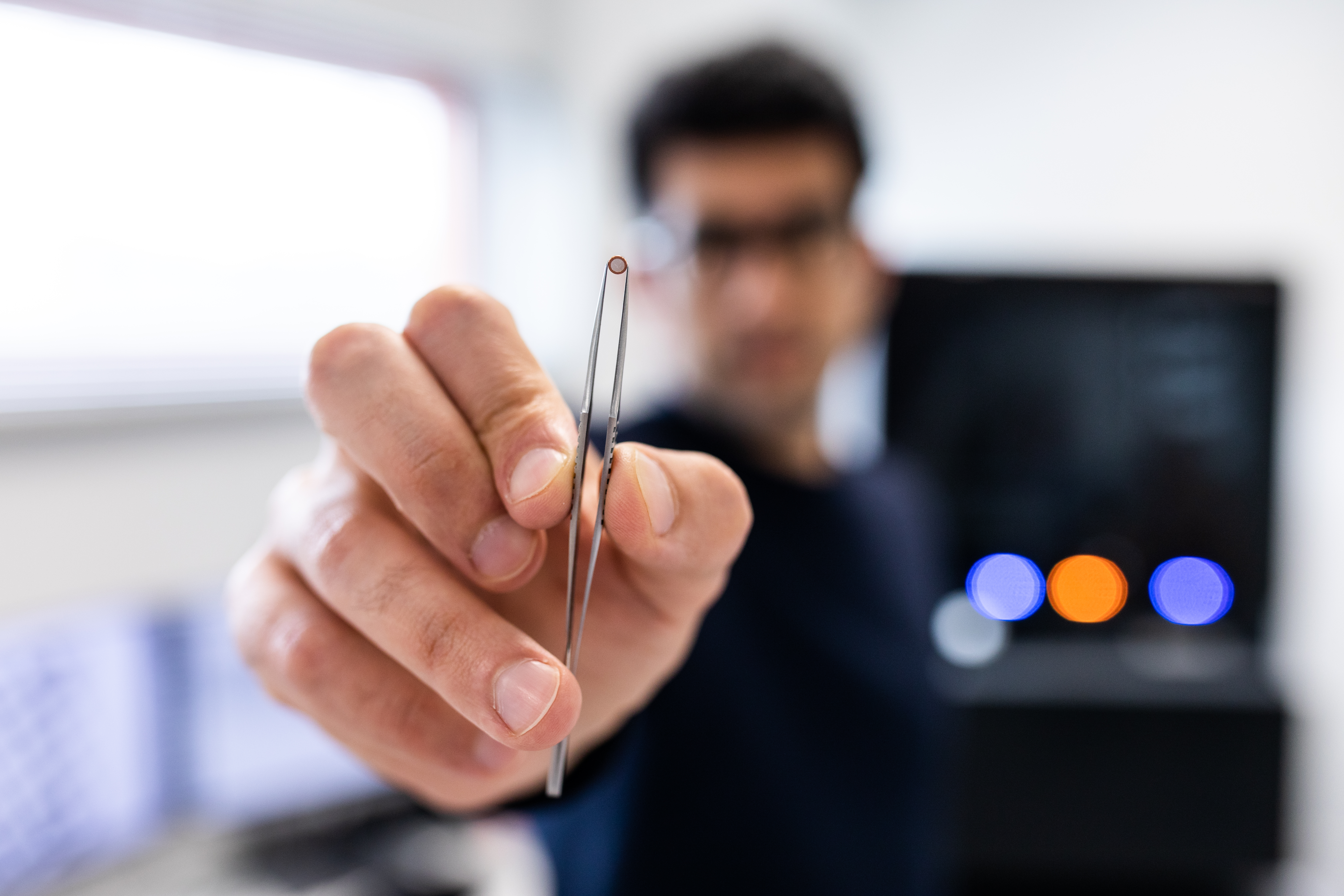
The material of the support mesh is pivotal for sample preservation and image quality. Copper grids have been the go-to choice due to their stability and conductivity. In certain cases, such as samples sensitive to copper ions, you might want to consider gold grids. Gold grids offer enhanced purity and biocompatibility, reducing the risk of contamination. The support mesh comes in various mesh sizes, with the most used grids ranging from 200 to 400 mesh. The mesh size dictates the level of sample support and space available for vitrified ice. Smaller mesh sizes provide better handling support, minimizing damage, but may limit your imageable area containing your particle of interest. In contrast, larger mesh sizes offer more imageable area but come with stability trade-offs. In the VitroJet, all grids are applicable, but the highest imageable area can be obtained with a low size mesh, e.g. 200. For making the right choice, consider the specific requirements of your sample and their potential impact on the material response.
Foil patterns: Tailoring support to your needs
The film covering the support mesh grid is of importance when there is a possibility of affinity with the sample, pattern or hole size. Additionally, the hole-to-carbon ratio needs to be considered when your sample can have affinity with the film layer. Larger holes give you large imageable area, but also a higher chance of beam-induced motion, specifically with thin layers which can be negatively affected by electron dosage. Furthermore, the choice of foil pattern can impact the distribution of holes and the ease of data collection. Other film types, such as Lacey grids, which contain a spider web-like film covering the support mesh, can offer advantages such as thinner ice thickness and enhanced contrast. However, the Lacey grids tend to be more fragile. For a first test, a grid with an intermediate hole size and distribution of 1.2/1.3 (1.2 µm hole size, 1.3 µm carbon in between each hole) is a good choice based considering possible beam-induced motion, ease of data collection and grid fragility.
The grid you select can make all the difference in your quest for near-atomic resolution, and your choices are crucial. So, dive in, explore, and advance your research. Your next breakthrough may be just a grid away. For a more in-depth look at this grid selection procedure and the newest innovations considering grid types, read on in our most recent application note!
About the author
Maaike Schotman is a cryo-EM enthusiast from the Netherlands with a background in biomedical engineering. She is passionate about uncovering the secrets of the structural protein world through cryo-EM and sample preparation. Beyond the lab, she enjoys bird watching and exploring the wonders of nature.